1898
The beginnings of radioactivity
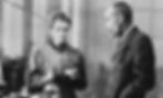
In 1898, Henri Becquerel, Pierre Curie & Marie Curie discovered the first radioactive elements, Polonium and Radium.
1932
Development of the first American cyclotron
Ernest O. Lawrence and Stanley Livingston created the first cyclotron put into service at the University of California in Berkeley.

1944
Samuel Curran and the modern scintillation counter
The first electronic scintillation counter was invented by Sir Samuel Curran whilst he was working on the Manhattan Project at the University of California at Berkeley.
1952
The first human positron imaging system
Gordon L. Brownell developed the first positron imaging system used for the human brain at the Massachusetts General Hospital. The Gordon Centre for Medical Imaging in MGH was named in his honor.
1963
David E. Kuhl and the Tomographic Imaging technology
During his residency at the University of Pennsylvania, David E. Kuhl, in collaboration with Roy Edwards, developed a tomographic scanner. Their approach is now used in PET scanners.
1975
The first ever PET scanner
After develpping a prototypical PET scanner, Michael E. Phelps, Edward Hoffman and and Michel Ter-Pogossian created a whole-body PET scanner with hexagonal detectors. It was the first clinically applicable PET scanner and was used for animal and human studies at the Washington University School of Medicine.

1976
Fluorodeoxyglucose first administration
It was not before 1976 that FDG was first administrated into a patient's body; it was performed by Abass Alavi and his team at the University of Pennsylvania.
The first PET scanner was installed at the University of California, Los Angeles (UCLA). Now Director of Nuclear Medicine at UCLA, David E. Kuhl conducted the first FDG-PET study.
Although PET imaging was very useful in its early stages, poor resolution significantly affects its performance and usefulness in both clinical and preclinical studies. In order to produce useful images, it soon became clear that a better spatial resolution was needed.
Storytime.
The History of PET.
1980s
The beginnings of what became the LabPET technology
In the early 80s, Professor Roger Lecomte undertook a major challenge to create a scanner that would allow the use of PET to conduct preclinical studies on small animals. Back then, it was impossible to image small animals with existing PET scanners since spatial resolution was too coarse to resolve internal organs or grafted tumors in mice or rats.
Dissection techniques were mainly used for several applications now performed with PET scanners, which limited the ability to understand disease progression and the effectiveness of developed treatments.

1990s
The first prototype
In 1995, after more than a decade of active research and dozens of scientific publications, Roger Lecomte and his team succeeded in setting up a first prototype demonstrating the proof of concept and potential of the technology developed for small animal imaging, triggering the molecular imaging revolution.

1990s
The issues of LabPET 0
In its early days, the LabPET scanners required a lot of space to operate. During the installation, 2 rooms were required to house the data acquisition electronics and the computers.
This was not unique to LabPET since at that time, data acquisition technologies relied on printed circuit boards with discrete components and required a lot of space and power. This aspect of the design did not affect their performance, but was making the scanners costly and cumbersome.

2000s
The LabPET I and its commercialization
After few years of R&D and engineering developments, the team finally came up with the next generation of the LabPET, launched in the early 2000s. In addition to reducing the space occupied, the highly integrated technology developed for the scanners was more efficient, achieved a better spatial resolution and was better adapted to preclinical imaging of small animals.
The first generation of commercial preclinical scanners called LabPET I had many scientific and commercial successes since the devices were very reliable, efficient and allowed many new scientific discoveries.

2010s
Development of the
LabPET II
After the great success of the LabPET I, the team developed the second generation of the LabPET to push the spatial resolution to the physical limit achievable in PET. In order to produce very high-quality images, all efforts were put into developing a modular detector offering the best possible resolution reaching 0.76 mm in the mouse version.
The LabPET II scanner technology for PET therefore includes a set of patents, innovations and manufacturing processes that have been developed for more than 30 years at the University of Sherbrooke. The technology is based on a matrix of pixelated detectors and has parallel electronic channels at an unprecedented level of integration (~ 70 channels/cm²), which allows the best spatial resolution in the world (0.8 mm isotropic or 0.5 µL) to be achieved, without distortion in the images, and at a very high maximum count rate. This high level of accuracy and the high quality of the images obtained have a direct impact on the research results. This type of scanner is used in specialized research centres around the world to conduct studies on animals (mainly mice, rats, rabbits and macaques) in order to better understand both normal physiology and human diseases, to develop new drugs or to test the effectiveness of new treatments against cancer, for example.

2010s
The first results of the LabPET II
The performance evaluation of the LabPET II technology was methodically initiated by a PhD student at the University of Sherbrooke using NEMA protocol, which are internationally recognized scientific standards for measuring the performance of PET scanners and are also used during the final acceptance of scanners after their installations. These initial tests made it possible, among other things, to quantify the 3 main important PET parameters: spatial resolution, sensitivity, and counting rates.
The actual measured spatial resolution is 0.74 mm in the middle of the scanner’s field of view, surpassing all other PET technologies currently available on the market. The sensivity reaches 3.3 % (with an energy window of 250-650 keV), and the effective metering rate (NEC) is 250 kcps at 70 MBq. Finally, structures and details never observed before in preclinical PET imaging were highlighted on mouse images obtained with the scanner, demonstrating the drastic improvement in quality that the technology offers.


Meanwhile.
The History of IR&T.